- AdventHealth University

Medical imaging technologies continue to evolve, presenting new applications in diagnosing and treating diseases. New medical imaging technologies promise to lead directly to improved patient outcomes. Breakthrough innovations, such as the ones below, are reshaping the medical imaging field:
- Artificial and augmented intelligence
- Augmented and virtual reality combined with 3D imaging
- New approaches to nuclear imaging
- Portable and wearable scanners
Chapter 1: History of Medical Imaging technology
In the 126 years since the accidental discovery of X-rays by Wilhelm Rontgen in 1895, medical imaging technology has revolutionized nearly all aspects of healthcare.
- Ultrasound imaging: In the 1960s, medical professionals began using ultrasound imaging to search for tumors and other growths without performing surgery.
- Magnetic resonance imaging: First developed in the 1970s, MRI provides a sharp noninvasive view of the body’s internal organs and tissues without the use of radiation.
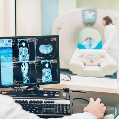
- Computed tomography: CT arrived about the same time as MRI, offering cross-sectional images of the body.
- Positron emission technology: PET also dates back to the 1970s, but in the 1990s, researchers combined the technology with CT scans to diagnose cancer using three-dimensional images. [Doctors Imaging, Mayo Clinic I]
Chapter 2: Recent Advances in Medical Imaging Technology
Medical scientists are adapting artificial intelligence (AI), virtual reality (VR), wearable computers, and other technological advances to improve healthcare. Computers can simulate real-world situations in ways that provide humans with new insights and diverse perspectives that go beyond human sensory perception.
- Adoption of AI and VR
While AI-based imaging operates without direct human intervention, augmented intelligence enhances human diagnostic capabilities to provide many of the benefits of AI, but with human reasoning combined with automated reasoning. Virtual reality and augmented reality technologies benefit radiology training by creating immersive environments that make complex anatomical structures easier to conceptualize.- DeepMind: Google’s AI system helps to reduce false results in breast cancer screening. [Google
- ProFound AI: iCad’s innovation applies machine learning and other AI techniques to enhance the accuracy of tomosynthesis imaging used to detect breast cancer. [iCad]
- Cardiac MRI segmentation and analysis model: Developed by Intel and Siemens, this model promises to deliver real-time diagnosis of cardiovascular disease. [Intel]
- Augmented intelligence: Automating some of the more mundane diagnostic processes provides a way to improve communication between radiologists and oncologists, for example.
- True 3D: EchoPixel’s virtual reality platform assists surgeons as they prepare to perform procedures to treat congenital heart defects. The patient-specific surgical views provided by the True 3D software reduce the likelihood of encountering unexpected conditions during surgery. [EchoPixel]
- Nuclear Imaging Advancements
Nuclear imaging functions by introducing radioactive material inside the patient’s body. A scan using a gamma camera senses the material wherever it concentrates in the body. The tests can detect thyroid disease, gallbladder disease, and many other common health conditions. Advancements in nuclear imaging technology are broadening the reach of the procedures.- Amyloid PET imaging: Showing tremendous promise in the early detection of Alzheimer’s disease, amyloid PET imaging demonstrates accuracy in noninvasively identifying and managing neurocognitive deficit. [Diagnostics]
- EXPLORER Total-Body PET Scanner: This groundbreaking tool features a much higher level of sensitivity than conventional PET scanners and is able to perform a full scan of the human body in a single acquisition in 99% of cases. [Journal of Nuclear Medicine]
Chapter 3: A Bright Future for New Medical Imaging Technologies
Innovative medical imaging technologies promise to improve patient outcomes in a range of areas. For example, mobile imaging equipment can monitor and diagnose elderly patients without an in-person medical appointment. Wearable medical devices with imaging components make it simpler and more convenient for patients to report their vital statistics.
- A mobile magnetoencephalography (MEG) scanner can measure brain activity while people are moving naturally, such as making gestures, stretching, or playing sports. This can make the diagnosis of conditions such as epilepsy more accurate by identifying the exact location in the brain that is the source of epileptic seizures. [Discover]
- An MRI glove can produce detailed 3D anatomical images of a hand in motion as it performs various activities, such as grasping or flexing, to better model the soft-tissue mechanics of the patient’s hand. [U.S. National Institutes of Health]
- Radiologists and diagnosticians will increasingly use 3D printing to educate patients and assist in the training of surgeons and other medical professionals. The 3D models give patients and students a clearer understanding of diseases and procedures to correct them.
- Interventional radiology will extend the use of advanced medical imaging beyond diagnostics to treatment and prevention. For example, geniculate artery embolization allows knee inflammation due to osteoarthritis to be treated via microcatheters and wires inserted vascularly. [Diagnostic Imaging]
- Researchers have developed techniques for noninvasively imaging and manipulating biological processes that occur deep inside the body. Genetically encoded reporters for ultrasound create imaging agents based on gas vesicles, which are hollow protein nanostructures derived from buoyant microbes. This allows microorganisms within a host organism to be identified and studied in previously inaccessible tissues. [Nature I]
- Researchers have also developed MRI reporter genes that allow molecular processes in living cells to be monitored noninvasively, which gives researchers and caregivers a better understanding of cellular pathology and therapy. [Nature II]
- 3D phase microscopy, being developed by a team led by scientist and computer imaging researcher Laura Waller, is another technique for imaging biological processes that existing technology can’t model.
- The new approach to quantitative phase imaging (QPI) measures optical path length changes to determine 3D refractive index values within cells. This allows researchers and clinicians to create shape and density maps of cells, making it easier to study subcellular structures. [Nature III]
- An LED array microscope in development replaces a traditional microscope’s lamp with an LED array for brightfield, darkfield, phase contrast, super-resolution, and 3D phase imaging via computational illumination techniques. [Nature IV]
Conclusion
Fast and accurate diagnosis is key to preventing and treating the maladies that threaten our health and well-being. With each new advancement, medical imaging technology becomes more important to detecting and addressing nearly all of the diseases that humans risk developing or contracting. Medical imaging professionals play a key role in the use of technological innovations in medical imaging to promote and protect our health.
Sources
https://blog.definitivehc.com/future-trends-in-medical-imaging-2019
https://deepmind.com/research/publications/International-evaluation-of-an-artificial-intelligence-system-to-identify-breast-cancer-in-screening-mammography
https://www.dicardiology.com/article/emerging-trends-medical-imaging
https://www.discovermagazine.com/mind/mobile-meg-will-new-technology-change-neuroscience
http://www.doctorsimaging.com/the-history-of-medical-imaging/
https://echopixeltech.com/applications.html
https://www.icadmed.com/profoundai.html
https://www.itnonline.com/article/emerging-trends-radiology
https://www.ncbi.nlm.nih.gov/pmc/articles/PMC6627350/
https://www.ncbi.nlm.nih.gov/pmc/articles/PMC6424228/
https://newsroom.intel.com/news/siemens-healthineers-intel-demonstrate-potential-of-ai-real-time-cardiac-mri-diagnosis/#gs.uo4jv3
https://www.mayoclinic.org/tests-procedures/mri/about/pac-20384768
https://www.mayoclinic.org/tests-procedures/pet-scan/about/pac-20385078
https://www.nature.com/articles/nmeth.4621
https://www.nature.com/articles/s41598-020-77576-z
https://www.nature.com/articles/s41598-019-55330-4
https://www.nature.com/articles/s41598-019-43845-9
https://www.ramsoft.com/the-history-of-positron-emission-tomography/


