|
Authored by:
- AdventHealth University

If you’ve never had an ultrasound exam, you might feel anxious about it, but the procedure doesn’t have to be stressful. The key to feeling confident is preparation: learning about the purpose of ultrasound scans, the basic procedures of an ultrasound exam, and issues you may want to discuss with your ultrasound technologist or other healthcare provider. By gaining a basic understanding of these topics, you’ll know what to expect, and discover areas where you might want to do additional research.
The Importance of Ultrasound Procedures
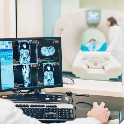
Diagnostic ultrasound, or sonography, gives healthcare providers the ability to see inside the body — viewing organs, tissue, and vessels — without making an incision. A key benefit of ultrasound is that it doesn’t involve radiation, so it is considered safer than an X-ray exam.
Common Uses for Ultrasound
Ultrasound enables healthcare providers to monitor and diagnose a wide variety of conditions and diseases.
Prenatal Care
While sonograms can be performed on many parts of the body, one of the most familiar ultrasound exams are pelvic scans of pregnant mothers. Providing images of the reproductive organs and the baby, prenatal ultrasounds are used to monitor infant growth and detect medical issues experienced by the baby and mother. Resources for pregnancy-related ultrasound information are listed below.
Ultrasound During Pregnancy: The March of Dimes, the nonprofit that works for the health of mothers and babies, details the different ultrasound technologies used during pregnancy and the different applications and screenings that can be performed.
Ultrasound Exams: This resource, from the American College of Obstetricians and Gynecologists, addresses frequently asked questions about ultrasound exams.
Cancer Diagnosis and Treatment
Ultrasound exams are used to help detect certain types of cancer, including breast and prostate cancer. Ultrasound technology is also used to guide needles for biopsies and tumor treatment.
Prostate/Rectal Ultrasound: This resource from Johns Hopkins Medicine details the procedure of prostate or rectal ultrasound tests, including the steps, risks, and preparatory measures.
Ultrasound: Breastcancer.org explains the use of ultrasound as a complementary procedure to other screening tests.
Ultrasound for Cancer: The American Cancer Society provides information on the use of ultrasound exams to detect tumors and aid in biopsy procedures.
Ultrasounds are also used to assess joint inflammation, diagnose gallbladder disease, check thyroid glands, and evaluate blood flow and metabolic bone disease, according to the Mayo Clinic. Additionally, ultrasound is used to assess cardiac and vascular conditions, often serving as a screening method in the prevention of cardiovascular disease.
What to Expect
Understanding how ultrasound scans work and what to expect during an ultrasound examination can alleviate the natural anxiety associated with the procedure.
How Ultrasound Works
The term ultrasound refers to high-frequency sound waves that are used to visualize soft tissue. Ultrasound scans are performed by trained technologists called sonographers. The sonographer operates a transducer, a device that generates and senses ultrasound energy. Commonly taking the form of a handheld wand, transducers are placed either on the surface of the patient’s body or internally. Sound waves bounce back and forth from the transducer, gathering data that a computer compiles to generate an image of the area being scanned.
Ultrasound: This resource from the Mayo Clinic provides an overview of the reasons ultrasound tests are performed and what patients can expect during the procedure.
Ultrasound Imaging: This resource on ultrasound imaging from the Food and Drug Administration (FDA) covers a basic description of the technology, its uses, benefits and risks, and information for patients, healthcare providers, and ultrasound practices.
Understanding Sonography: The Society of Diagnostic Medical Sonography provides information ranging from the history of ultrasound technology to the various conditions that can be detected and diagnosed with ultrasound.
Procedure
For external ultrasounds, the sonographer applies a water-based gel to the area that is being scanned. The gel allows the transducer to move easily back and forth to capture an image. The gel also prevents air pockets that could block the sound waves that are being used. External ultrasound scans are usually not painful, and patients typically experience no discomfort. (One exception is pregnant women who are asked to maintain a full bladder for the exam.)
For internal ultrasounds, the transducer is placed in a man’s rectum or a woman’s vagina, if the urinary system or reproductive organs are being evaluated. For certain examinations of the digestive system (esophagus, chest lymph nodes, stomach) or heart, an endoscope is used that has a light and ultrasound device attached to the end. The endoscope is inserted through the mouth. Patients usually receive medication prior to internal ultrasounds to reduce pain and discomfort.
Patients are asked to lie on their back for internal and external pelvic procedures, or on their side for endoscopic and cardiac procedures. In some cases, patients may receive a contrast agent via injection that makes ultrasound images appear clearer. A typical ultrasound exam lasts 30 minutes to an hour.
Endoscopic ultrasound: This overview of endoscopic ultrasound provided by the Mayo Clinic explains why the procedure is performed, steps to prepare, and associated risks.
Ultrasound – Pelvis: RadiologyInfo, a website developed by the Radiological Society of North America and the American College of Radiology, offers a detailed description of pelvic ultrasounds, including transabdominal, transvaginal, and transrectal procedures.
Results
The timing of ultrasound results depends on the procedure and purpose of the exam. For example, it is common during prenatal scans for doctors to discuss the results during the exam. For purposes such as tumor identification, the doctor may require more time to review the images. It may take a few days to receive results, and the doctor may schedule a meeting to discuss exam findings or schedule a follow-up for more testing. Depending on the exam results, the physician may order a different type of scan (CT, MRI) or take a biopsy sample (tissue sample).
Although there is nothing to fear from an ultrasound exam, it is normal to experience some anxiety about the results of any diagnostic tests. Asking a healthcare provider to address any questions you have before you leave the medical facility can help reduce stress and concern.
Getting a Second Opinion: The American Cancer Society provides advice for talking to your doctor about getting a second opinion and steps to do so.
How to Ease Worry When Waiting for Medical Test Results: This article from Patient.info outlines some strategies for addressing anxiety related to test results.
Preparing for an Ultrasound
Ultrasound scans are relatively simple procedures, but proper preparation can improve the experience. The following lists of tips and questions address basic ultrasound preparation.
Tips
Although the required preparation for ultrasound exams is generally not extensive, one pre-exam rule should be addressed carefully: follow your healthcare provider’s instructions regarding consumption of food and water. Patients may be asked to refrain from eating for several hours before a scan is performed. This is because undigested food can block sound waves. For other scans, a patient may be instructed to drink large amounts of water and hold their urine, because a full bladder makes it easier to visualize pelvic organs.
The following are other preparation tips to consider.
- Wear loose-fitting, comfortable clothing. Depending on the type of scan, you may be asked to remove some clothing or put on a hospital gown.
- Refrain from wearing jewelry, because you may be asked to remove it.
- Let your doctor know if you have a latex allergy, so they will be sure not use a latex-covered probe.
- Tell your doctor about any over-the-counter (OTC) or prescription medications or herbal supplements you’ve been taking.
Questions
Patients should always talk to their healthcare provider if they have any questions about a procedure. Sonographers and the doctors and medical specialists they work with should communicate necessary information before, during and after the exam, but questions may still arise. During prenatal exams, for example, parents may have specific questions about development of a baby’s organs or the mother’s health. More generally, patients should make sure they understand the answers to the following questions.
- What is the test for and what do the results mean?
- What risks are associated with the test, and are there potential side effects or complications?
- Who will perform the test and what are their qualifications?
- When will results of the test be available?
- Who will provide answers if there are questions about the test or results?
Fetal Ultrasound: Provided by Stanford Children’s Health, this resource includes a more comprehensive list of information that a patient should gather before a test or procedure is conducted.
Preparation Is Key
Diagnostic ultrasound is a proven method for safely and effectively gathering critical imaging data. It poses minimal discomfort for patients, but it does have its limitations. By learning more about ultrasound technology prior to an exam, patients can reduce their anxiety about the procedure and instead focus on their health.
Additional Resources:

