|
Authored by:
- AdventHealth University

Imaging technologies, such as ultrasounds and mammograms, are used in women’s health to help diagnose conditions and improve health outcomes. There are a wide variety of women’s health conditions that require or can benefit from imaging tools. These situations include a pregnant woman receiving an ultrasound, or a digital mammogram helping to detect breast cancer.
Diagnostic radiology and interventional radiology are vitally important to the medical field and regularly save the lives of many women. It’s clear that tools such as ultrasounds and x-rays help doctors, technologists, and medical professionals improve women’s health.
Radiologic and MRI technologists who work in the health field can have rewarding careers centered around imaging focused on women’s health. Technologists who are interested in pursuing a career in imaging for women’s health may wish to explore the Bachelor of Science in Imaging Sciences offered at AdventHealth University Online.
What Is Imaging for Women?
When the German physicist Professor Wilhelm Röntgen performed the first x-ray in 1895, he didn’t know exactly what he had accomplished or how it would provide lasting benefits for the medical field. Soon after, scientists began developing x-ray technology. Their development efforts included experimenting with sonar and sound waves, which led to ultrasound machines.
Ultrasounds began to grow in prominence throughout the 1960s. From the beginning, doctors used ultrasound technology to examine patients for tumors. Physicians continue to use ultrasound technology today when searching for tumors and cancer. They also perform ultrasounds on women during pregnancies to monitor fetal development.
After the advent of ultrasounds, healthcare imaging for women continued to evolve. Dr. Raymond Damadian created the first magnetic resonance imaging (MRI) machine, and Sir Godfrey Hounsfield created the first computed tomography (CT) machine in the 1970s. These complex imaging technologies have continued to advance over the last fifty years and have become necessary tools in the medical field.
While imaging technology is used on all patients to diagnose the cause of their symptoms, health imaging for women has grown in importance. It has helped diagnose a variety of conditions, from breast cancer to osteoporosis. With x-rays, such as mammograms, doctors have been able to more successfully identify tumors and breast cancer, and begin treatment on women at earlier stages. Additionally, ultrasounds are known to produce high-quality images of fetuses at different stages of growth.
As healthcare knowledge continues to advance, there are additional applications of imaging technology in promoting health and wellness. Many female patients also benefit from lesser-known technologies that are used in imaging for women’s health, such as Positron Emission Mammography (PEM), a type of nuclear medicine scan that provides early detection of breast cancer in high risk patients. Women also benefit from Bone Mineral Density Exams, a type of x-ray exam that helps screen for and identify osteoporosis.
Types of Women’s Health Imaging
As technologies have advanced over the last several decades, particular imaging technologies have demonstrated many benefits for treating conditions and promoting wellness for women.
Mammograms
Mammograms can be performed for routine breast screening or to diagnose a specific concern within the breast. Mammographers work with patients who have been referred by their doctor and perform mammograms in hospitals, doctor’s offices, outpatient imaging facilities, and even mobile mammography units. According to the Centers for Disease Control and Prevention (CDC), 65.3% of women over forty years of age have had a mammogram in the last several years. Mammographers play an important role in discussing the procedure with patients and following specific imaging protocols to obtain the necessary images.
Ultrasounds
Ultrasounds make use of sound waves to create images of internal organs and tissues. This type of imaging technology does not require radiation and is most commonly known for helping provide healthcare imaging for women during pregnancy. Ultrasound technologists, also known as sonographers, can perform ultrasounds on pregnant women to help monitor growth, determine gender, and identify the position of the fetus in the womb. Ultrasounds are also used in gynecological exams to detect cervical and ovarian cancer as well as for breast imaging. Additionally, ultrasounds are used for many other conditions, and can help physicians identify the cause of pain in various parts of the body, such as the kidneys and the thyroid.
Bone Mineral Density Exams
This imaging technology uses radiation to provide information about bone density and the amount of calcium in a patient’s bones. The test can help screen for osteopenia and osteoporosis, enabling physicians to provide early treatment if indicated. Bone mineral density scans, also known as DEXA scans, can help doctors accurately measure the levels of bone density, make predictions about patients’ conditions, and ascertain whether or not osteoporosis medications are working effectively. Doctors typically recommend bone mineral density exams for women who are older than sixty-five or are over fifty years of age and have broken a bone.
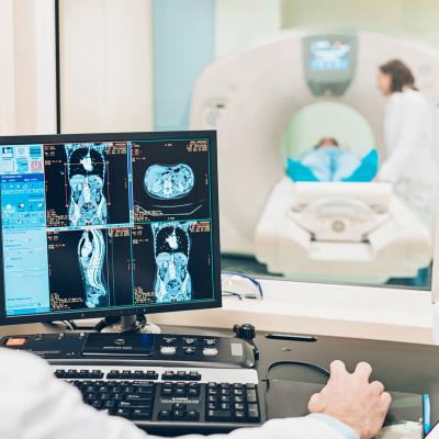
MRI Scans
MRI scans can be used in breast imaging for women after mammograms have been completed and if doctors need to perform further diagnosis. This imaging technology allows for a combination of high-resolution images from various angles, providing physicians with enhanced information about tissue type and density. In some cases, fetal MRI scans can be performed if ultrasound alone is not able to provide a diagnosis.
Careers in Imaging for Women
Imaging technologies are a vital component of the medical field and continue to support women’s health in a variety of important ways. There are several different specialized careers in radiologic sciences, including those for mammographers, diagnostic medical sonographers, and MRI technologists.
Many individuals begin these careers by earning an associate’s degree and receiving state licensure and/or national credentials. While pursuing a bachelor’s degree, imaging technologists can expand their understanding of how to use imaging equipment, perform advanced procedures, manage imaging departments, and navigate regulatory standards in radiology. Earning a bachelor’s degree can also help technologists advance their careers in health imaging for women.
According to the U.S. Bureau of Labor Statistics (BLS), the job outlook for careers in imaging and radiologic technology are projected to grow 9% between 2018-2028, which is faster than most careers. The latest data reveals the annual median salary for radiologic technologists is $59,520 and the annual median salary for MRI technologists is $71,670.
The employment outlook is also positive for sonographers, with jobs projected to grow 14% between 2018-2028. According to the BLS, sonographers make an annual median salary of $67,080.
Advance Your Imaging Career
Imaging for women’s health takes many forms, through ultrasounds, MRI scans, mammograms, bone density tests, and other imaging technologies. Students and professionals who are focused on imaging careers can develop their skills and expand their career horizons by pursuing a bachelor’s degree in the field. Students who want to advance their imaging careers can pursue AdventHealth University Online’s Bachelor of Science in imaging Sciences program to prepare for successful careers in the medical field and make positive impacts on women’s health.
Recommended Readings
How to Start a Sonography Career: Steps, Tips, and Resources
Safety Concerns for Sonographers
Sources:
BIDMC, “Ultrasounds During Pregnancy: How Many and How Often?”
Centers for Disease Control and Prevention, Mammography
Doctors Imaging, “The History of Medical Imaging”
Health Imaging, “USPSTF Updates BRCA Cancer Screening Recommendations”
MedlinePlus, “Imaging and radiology”
National Osteoporosis Foundation, “Bone Density Exam/Testing”
NPS Medicinewise, Imaging Explained
Science Direct, Imaging Technique
The Bureau of Labor Statistics, Radiologic and MRI Technologists

