|
Authored by:
- AdventHealth University

A patient with swelling and pain deep in one leg receives an order to get an MRA scan. A patient who’s having problems with leg movement after a fall receives an order to get an MRI. Same areas of the body, same directive to get an imaging scan. But different acronyms — and different tests.
MRA vs. MRI: What’s the difference?
MRI, or magnetic resonance imaging, uses radio waves, a magnetic field, and a computer to create images of the inside of the body. MRA, or magnetic resonance angiography — sometimes called a magnetic resonance angiogram — is a magnetic resonance procedure that zeroes in on the blood vessels.
As their similar names suggest, MRAs and MRIs are closely related. In fact, MRA is a type of MRI. Both procedures allow medical professionals to examine the inside of the body without making a single incision, but their processes and uses differ.
In the case of the two patients experiencing different problems in the same area, an MRA would produce images that would show blood clots that could be causing swelling and pain deep inside the leg. An MRI would provide images showing damage to tissues or ligaments that occurred as a result of the fall, causing mobility issues.
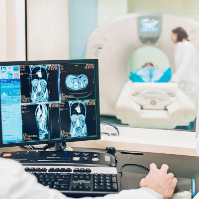
Imaging technologists can pursue a bachelor’s degree that allows them to focus on specific types of imaging modalities, including MRIs.
MRA vs. MRI: Definitions
MRAs and MRIs are diagnostic tools used to view organs, bones, or tissues inside the body. Developed more recently than X-rays, which use radiation and produce less detailed images, MRAs and MRIs rely on a strong magnetic field to provide a more defined picture.
Sometimes, MRAs and MRIs also use contrast media administered intravenously to allow for greater differentiation of structures in the images produced by the scan.
What Is an MRI?
First performed in the 1970s, MRI procedures capitalize on the body’s high water content. Many health issues can change the makeup of that water, and MRI images show changes in fat and water molecules to help healthcare professionals diagnose problems.
During a typical procedure — generally conducted at a hospital or diagnostic imaging center — the patient lies on a table that slides into the tunnel-like machine that performs the scan. Some imaging facilities offer machines that are more open, which can be helpful for people who are uncomfortable in confined spaces.
An electric current in coiled wires creates a temporary magnetic field, and the machine sends and receives radio waves to create digital images of the area of the body being examined. A computer uses these images of specific locations in the body to create multidimensional pictures.
What Is an MRA?
An MRA is sometimes used in conjunction with an MRI. MRAs evolved from MRIs in the 1980s to provide a more thorough look at the body’s major arteries. MRAs apply the MRI principle and technique to focus specifically on the blood vessels.
The images an MRA produces show parts of the 100,000 miles of tiny and complex pathways that carry blood through the body. Those images help physicians gauge how quickly the blood is traveling and identify obstructions.
MRA vs. MRI: Similarities and Differences
While MRAs and MRIs employ the same concept and process, the areas they examine and the images they provide can be different. Following is a look at MRA vs. MRI, examining their similarities and differences.
How Are MRAs and MRIs Similar?
MRAs and MRIs use the same type of machine. They require the person undergoing the procedure to lie flat and still, usually wearing headphones or earplugs to protect against noise while in the machine. Both exams may require the patient to receive a dose of metal-based, paramagnetic contrast through an IV typically in the arm.
The procedures can take anywhere from around 15 minutes to more than an hour. The MRI technologist performing the test communicates with the patient via intercom at the same time. MRAs and MRIs produce images from inside the body. A radiologist interprets those images, advising the patient’s physician of the results to share with the patient.
For MRAs and MRIs, patients must remove jewelry and other metal objects. Other items that can interfere with these procedures include:
- Implants
- Pacemakers
- Joint replacements
- Tattoos
How Are MRAs and MRIs Different?
The main difference between MRAs and MRIs is the area of focus. MRIs produce images of organs and tissues in areas such as the brain, the heart, and joints. The procedure can examine parts of the body including:
- Muscles
- Bones
- Organs
- Tissues
MRAs’ focus is narrower. The exam offers a detailed look at blood vessels and how they’re functioning in different parts of the body, rather than focusing on the organs or tissue surrounding them. It also can show the rate of blood flow and circulation. While MRIs produce both 2D and 3D images, MRAs produce mainly 3D images.
MRA vs. MRI: Uses for Each Procedure
MRAs address vascular issues, identifying problems related to heart disease or stroke, for example. MRIs examine larger areas in the body to detect problems such as tumors or tears in soft tissue.
When Do Healthcare Professionals Use MRIs?
MRIs can help in diagnosing or ruling out a variety of diseases and conditions, including:
- Bone infections
- Arthritis
- Brain injuries
- Tumors
- Multiple sclerosis
- Inner ear or eye injuries
- Spinal cord damage
- Cancer
- Torn cartilage, ligaments, or tendons
So, when a patient is experiencing pain and mobility issues in one leg, as in the opening example, an MRI could help determine if the fall caused damage to cartilage, ligaments, or tendons.
When Do Healthcare Professionals Use MRAs?
Physicians order MRAs to measure whether blood is moving too fast or too slowly, often in cases of stroke, heart disease, or a blood clot. Blood that’s moving too quickly could indicate high blood pressure, while blood that’s too slow could indicate a blockage that might cause a heart attack. MRAs help medical professionals examine blood flow between the kidneys, brain, and legs.
In the prior example of a patient experiencing pain and swelling in one leg, a physician might call for an MRA to determine if a blood clot is causing the symptoms.
Advance Your Imaging Career With a Bachelor’s Degree
If you’re ready to learn more about MRAs vs. MRIs — and take the next step in your imaging career — explore AdventHealth University’s Bachelor of Science in Imaging Sciences Online. The program offers six specialty tracks, including an MRI Track, with leaders in the field sharing the latest findings and techniques.
You’ll prepare for potential leadership opportunities and workplace advancement on a flexible schedule and with multiple resources for support. Discover how AdventHealth University’s Bachelor of Science in Imaging Sciences Online program can help you achieve your career goals.
Recommended Readings
7 Patient Care Tips for Imaging Technologists
How to Become an MRI Technologist
Types of Medical Imaging: Technologies and Career Options
Sources:
AICA Orthopedics, “Why Might My Doctor Recommend an MRI?”
American Society of Clinical Oncology Cancer.Net, Magnetic Resonance Imaging
Doctors Imaging, “What Is the Difference Between MRI and MRA?”
HealthHearty, “MRI vs. MRA: The Subtle Differences You May Not Know”
Health Images, “The Difference Between an MRI and an MRA”
Johns Hopkins Medicine, Magnetic Resonance Angiography (MRA)
Radiology Key, “Magnetic Resonance Angiography of the Abdomen”
ThoughtCo., “A Guide to Magnetic Resonance Imaging (MRI)”
U.S. Food & Drug Administration, Magnetic Resonance Imaging (MRI)

